functional ultrasound imaging (fUS/fUSI) is a versatile technology to image brain activity
at high spatiotemporal resolution, with a large field of view and depth of field.
The fUSI can be used for preclinical and clinical neuroscience research.
Large-scale neural circuits imaging
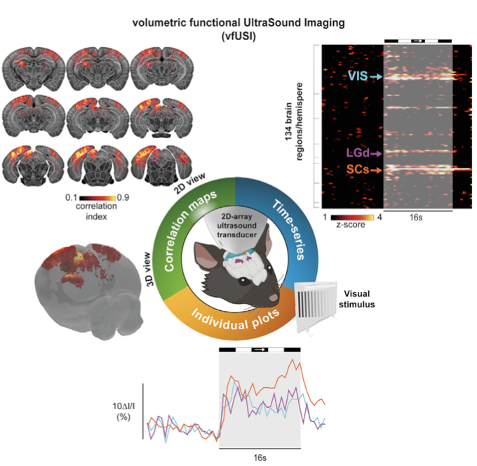
fUSI is a unique technology for decoding brain activity at mesoscopic scale offering unprecedented spatio-temporal resolution (100µm/100ms), large field of view (several cm) and depth of field (up to 6.5cm). fUSi does not require contrast agent and can be used in a large variety of animal models including rodents, non-human primates and even birds.
Costless alternative to fMRI to study brain pathologies
Get rid of the confounding effects of anesthetics
The fUSI technology allows both measurement of brain activity and blood flow in awake and chronic conditions. fUSI is efficient to track changes over time in various pathologies, including stroke, traumatic brain injury, vascular cognitive impairment (AD, PTSD, TBI), and more.
Complementary to in vitro research
Nowadays it is impossible to mimic the entire brain in a dish. fUSI is suited for a wide range of applications, including in vivo pharmacology studies. Visualize the effects of your active compound directly in the brain using functional activity, resting-state or dynamic connectivity mapping.
Fully compatible with electrophysiology and a wide range of techniques
fUSI is compatible with a large variety of technologies for the study of brain activity including 2-photons laser-scanning microscopy, electrophysiology with the IMEC Neuropixels, flex-polyimide and silicon probes, optogenetics, pharmacogenetics and more…
Ready for clinical research
fUSI is a versatile technology for imaging brain perfusion and activity non invasively in neonates and adults for image-guided procedures
TRANSDUCER FREQUENCY
SPATIAL RESOLUTION
TEMPORAL RESOLUTION
FIELD OF VIEW
PENETRATION
Latest Publications
This section presents the recent scientific publications and news using the fUSI technology
November 2021
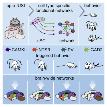
ARTICLE| VOLUME 109, ISSUE 11, P1888-1905.E10, JUNE 02, 2021 Optogenetic [...]
NEURORESOURCE| VOLUME 108, ISSUE 5, P861-875.E7, DECEMBER 09, 2020 A [...]
“fUSI is a breakthrough technology for real-time imaging of brain circuits dynamics at large scale”